Pediatric Entities in Sports Medicine: Most Common Sports Injuries for Children
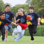
Every year in the United States, approximately 30 million children and adolescents participate in organized sports. An increase in pediatric sporting events also brings an increase in pediatric sporting injuries. There are nearly 3 million emergency department visits annually for pediatric-related sports injuries.
Medical personnel and athletic staff should become familiar with treating the pediatric sports patient. Skeletally immature athletes are unique because of the effects of growth on the musculoskeletal system. They are not only at risk for many of the injuries sustained by adults, but also may suffer injuries to the growth plates, apophyses, and joint surfaces. This article gives a general overview of the most common sports injuries seen in the skeletally immature athlete.
Salter-Harris Fractures
These are fractures localized to the growth plate of a bone in the skeletally immature patient. Salter-Harris fractures are classified into five different types. Type I fractures are fractures through the physis, or growth plate. On radiographs, a fracture usually will not be seen. It is common to see a widened physeal area between the metaphysis and epiphysis of the affected bone. Type II fractures involve both the physis and metaphysis. Type III fractures involve the physis and epiphysis. Type IV fractures involve the physis, metaphysis, and the epiphysis. Type V fractures are due to a compression injury of the physis. There will often be an absent physeal area between metaphysis and epiphysis on radiographs for Type V fractures.
The goal for treatment in Salter-Harris fractures is to obtain anatomic physeal reduction to avoid growth arrest. The risk of physeal growth arrest increases with Salter-Harris classification. Types I and II fractures usually can be treated with immobilization. In instances where there is fracture displacement, closed reduction may be necessary, followed by immobilization with casting. Rarely is internal fixation needed. Types III, IV, and V fractures usually require anatomic reduction with internal fixation to avoid growth arrest. These patients must be followed long-term to assess for growth problems.
Apophyseal avulsion fractures
The apophysis is a secondary ossification center and the site of tendinous attachment to bone. In the growing adolescent, muscle and tendon growth lag behind bone growth. This creates tension at the apophysis. A forceful contraction of muscle can then lead to an avulsion of the apophysis from its attachment site to bone. The most common area for this to happen is in the hip/pelvis. The hamstrings muscles of the posterior thigh attach to the ischial tuberosities, and the rectus femoris muscle (one of four quadriceps muscles) attaches to the anterior inferior iliac spine. Other areas of apophyseal avulsion fractures include the anterior superior iliac spine, greater and lesser trochanter of the femur, the tibial tubercle, and the base of the fifth metatarsal in the foot.
Athletes usually describe pain and a popping sensation during running, jumping, kicking, or stretching. There is usually tenderness over the affected apophyseal area and limited range of motion of affected muscle and limb. Radiographs usually show the avulsion. Treatment is often conservative. Rest, crutches, physical therapy, and NSAIDS are often the only treatment needed. However, if the displacement of the avulsed apophysis is greater than 2 cm, then open reduction and internal fixation may be needed.
Overuse injuries
Overuse injuries are types of secondary to repetitive sub-maximal loading that results in failure of connective tissues. Injury occurs when rate of fatigue exceeds rate of repair in these tissues. Some of the injuries this article will talk about deal with stress fractures, physeal, and apophyseal injuries.
Stress fractures are due to a failure of bone homeostasis. The rate of resorption occurs at a rate greater than that of formation. It is caused by four main mechanisms: repetitive muscle contraction leading to bony overload, increased bone stress following muscle fatigue, high repetition of low stress activity, or increased loads on hard surfaces. Stress fractures usually occur with sudden increases in activity intensity or multiple high intensity workouts without rest. Other factors related to stress fractures include poor arch support in footwear, improperly fitting footwear, and running on hard surfaces. Treatment for stress fractures includes discontinuation of inciting activity, rest, ice, limited weight bearing, and NSAIDS. Sometimes non-weight bearing activities and casting is required. Once the patient becomes pain-free, a slow resumption to activity is necessary. It may take 8-16 weeks or longer for return to full activity.
Apophyseal injuries are a common overuse injury and cause of pain in the adolescent athlete. Perhaps the three most common apophyseal injuries that occur are Osgood-Schlatter disease, Sindig-Larsen-Johannson disease, and Sever’s disease. Osgood-Schlatter disease is due to a traction apophysitis of the tibial tubercle. It is secondary to repetitive microtrauma. Most patients present between the ages of 8-15 and complain of tenderness, swelling, and a prominence at the tibial tubercle. Pain is usually worse with squatting, jumping, or kicking. No knee joint effusion will be seen. Plain film radiographs may show a prominence of the tibial tubercle. Treatment is mainly conservative. Ice, NSAIDs, activity modification, cho-pat, and knee padding are all modalities that seem to help. The disease itself is self-limited and usually resolves by the end of puberty.
Sindig-Larsen-Johansson disease is secondary to a repetitive microtrauma of the inferior pole of the patella. It occurs most commonly before the prepubescent growth spurt in males. Patients present with anterior knee pain localized to the inferior patella. Pain is worsened with running, jumping, ascending/descending stairs. Often times patients will have a limp and limited range of motion secondary to pain. Plain film knee radiographs will show elongation of patella with calcification located at the inferior pole of the patella. Treatment is self-limited. Ice, NSAIDS , and modification of activity all seem to help. Spontaneous resolution typically occurs about 12-18 months after onset of symptoms.
Sever’s disease, also known as calcaneal apophysitis, is a traction apophysitis located on the heel of the foot. It is most common in pediatric athletes age 9-14. It is bilateral in about 60-80 percent of cases. Patients present with calcaneal tenderness near apophysis, and heel pain worsened with activity. Calcaneal plain film radiographs may show sclerosis or fragmentation. However, most of the time the only thing seen will be an open growth plate. Treatment is self-limited. Heel cups, heel cord stretching, NSAIDs, ice, and activity modification are good conservative treatments.
Little leaguer’s shoulder is secondary to an overuse injury of the physis at the proximal humerus of the shoulder joint. It is also described as an osteochondrosis of the proximal humeral physis or a stress fracture of the proximal humeral epiphyseal plate. It typically occurs in males from age 12-15 years. It is highly associated with the quantity of pitches thrown in the young adolescent. Patients present with pain localized to the proximal humerus during the act of throwing. There is usually no inciting event, and the onset of pain is gradual. Playing ability and velocity of throw diminish with pain. Physical exam usually shows weakness in external rotation of shoulder, pain with resisted internal rotation, and normal strength and range of motion. Plain AP external rotation radiographs of the shoulder will show widened and irregular proximal humeral physis, metaphyseal fragmentation, and /or sclerosis of the metaphysis. Treatment usually involves rest from throwing for at least 6 weeks to 3 months. After this period of rest, the patient must then complete a throwing program before returning to full activity.
Little leaguer’s elbow (LLE) is an overuse injury of the physis of the medial elbow. It is secondary to valgus throwing stress of the medial elbow. It can cause a medial epicondyle apophysitis, medial epicondyle avulsion fracture, or osteochondrosis of the capitellum (Panner’s disease). It is most common in boys aged 9-14 years. Injuries on medial elbow primarily occur during the acceleration phase of throwing. Physeal injury of LLE usually presents with tenderness over the medical epicondyle. Radiographs are typically normal, but may reveal widening of medial epicondyle epiphysis. Treatment is mainly rest and initiation of throwing program after rest period. Medial epicondyle avulsion fractures usually present after forceful throwing. Patients usually describe a pop in the medial elbow followed by persistent pain. Radiographs will show an avulsion of medial epicondyle from medial elbow. If nondisplaced, patients are usually placed in a cast for 2-3 weeks and gradually begin return to play activity. If the fragment displaced is greater than 5 mm, then open reduction and internal fixation may be needed.
Panner’s disease usually occurs in pediatric patients 4-8 years of age. Patients typically present with painful throwing and tenderness at the radiocapitellar joint. They may have a 10-20 degree flexion contracture at the elbow. Early detection is crucial as progression of disease can be prevented with activity change. Plain film radiographs of elbow may show loose bodies or an irregular ossification center. MRI is helpful in defining the lesion. Treatment is usually conservative with cessation of throwing. Then an interval throwing program is begun once pain subsides and range of motion and strength are normal. Surgery is indicated for persistent pain, symptomatic loose bodies, or a locked elbow.
References
Adirim, Terry A., Cheng, Tina L. Overview of Injuries in the Young Athlete. Sports Med 2003. 33(1); 75-81.
Peck, David M. Apophyseal Injuries in the Young Athlete. American Family Physician. June 1995. 51(8). 1891-1895.
Soprano, Joyce V. Musculoskeletal Injuries in the Pediatric and Adolescent Athlete. Current Sports Medicine Reports. 2005. 4:329-334.