
Posterolateral Corner
Last Updated on November 7, 2023 by The SportsMD Editors
The posterolateral corner (PLC) functions to stabilize the knee against posteriorly and externally directed forces. Injuries to the PLC represent less than 2% of all acute ligamentous injuries to the knee. Though rare, this injury can have devastating consequences on athletic performance.
What is posterolateral corner of the knee?
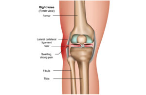 The PLC is comprised of multiple ligamentous and tendinous structures around the knee. A tendon represents connect tissue that attaches the muscle to the bone while a ligament is fibrous tissue that connects one bone to another. The PLC is located on the outside back (posterolateral) corner of the knee. Though many structures comprise the PLC, the main structures include the biceps femoris muscle and tendon (outside hamstring muscle), lateral collateral ligament, popliteus muscle and tendon, popliteofibular ligament, fabellofibular ligament, arcuate ligament complex, and posterolateral joint capsule.
Function of posterolateral corner
The multiple structures that make up the PLC function to stabilize the knee against certain directional forces. Specifically the PLC protects against posterior translation, external rotation, and varus rotation. A posterior directed force is when the tibia or shin bone is directed backward against the femur or thigh bone. An externally directed force is when the tibia is rotated outwardly or away from the midline of the body. A varus force is an inwardly directed force applied to the tibia from the outside.
Causes
Most PLC injuries occur as a result of trauma. 40% of these injuries occur from an athletic injury. A PLC injury can occur when the knee is hyperextended and a varus (or inwardly directed) force plus a rotation force is applied to the knee. This is commonly seen in football when a player is hit along the front inside of the knee with a force directed to the back outside of the knee. Although rare, noncontact injuries can occur when the knee is hyperextended and externally rotated. PLC can also occur from high energy trauma like a motor vehicle accident.
Other related injuries
It is rare for a PLC injury to occur in isolation. PLC injuries are commonly associated with anterior cruciate ligament (ACL) and posterior cruciate ligament injuries (PCL). The common peroneal nerve can also be damaged. This nerve supplies feeling to the top of the foot and helps extend the toes and ankle.
Classifications
PLC injuries can be classified based on the timing of the injury and based on the severity of the injury. From a timing standpoint, the injury is either acute or chronic. Acute injuries are generally less than 4 weeks old while chronic injuries are older. This is important because the age of the injury influences the surgical treatment options. Acute injuries can be repaired while chronic PLC injuries need to be reconstructed.
From a severity standpoint, PLC injuries can be graded on a scale from I – III. A grade I injury is a sprain to the PLC structures. There is no appreciable laxity or structural damage to the PLC. With grade II injuries, there is minimal laxity and injury to the PLC structures. Grade III injuries represent a complete disruption of the PLC structures with significant instability. The grade of the injury influences surgical intervention. In general, grade I and II injuries can be treated without surgery while grade III injuries require surgical intervention.
Diagnosis posterolateral corner injury
Taking a patient’s history and performing a physical exam will help your doctor diagnose a PLC injury. Patients with acute injuries will have pain along the back, outside (posterolateral) aspect of the knee. They may also have bruising there, along with neurologic findings if the common peroneal nerve is injured. Patients with long standing PLC injuries can have pain along the joint line both on the inside and outside of the knee. Neurologic findings may or not be present. The knee may also give way into hyperextension when walking up or down stairs or with twisting movements.
What tests might my doctor order?
Your doctor may order x-rays. This helps look at the overall alignment of the bone and assess for any fractures.
While an x-ray is useful for looking at the bone, an MRI will help visualize the ligaments and tendons around the knee. The MRI is also useful for evaluating other injuries within the knee.
Do I have to have surgery?
Treatment options for PLC injuries are based on many factors including the patient’s age, physical demands, activity level, athletic goals and expectations, severity of the injury, and other associated injuries. Grade I and II injuries can do well without surgery. They can be treated with crutches and a hinged knee brace for 4 to 6 weeks followed by physical therapy. With a grade III injury, the PLC structures are too damaged to heal on their own. This injury will require surgery.
Posterolateral corner surgery
Acute injuries (less than 3 weeks old) can be repaired. This involves sewing the ligaments or tendons back together or reattaching them back to bone. Results from an acute repair have better functional results compared to a reconstruction. When an injury is 4-6 weeks old, the damaged structures start to scar down. This can make a surgical repair difficult or even impossible. After 6 weeks it is recommended to reconstruct the PLC.
Reconstruction involves recreating the damaged PLC structures from other ligaments or tendons. Commonly the lateral collateral ligament, popliteus tendon, and/or the popliteofibular ligament are reconstructed. Currently, there is no gold standard for reconstructing the PLC.
Chronic PLC injuries can be associated with a malalignment or bowlegged deformity of the leg. If this is present, an osteotomy or realignment of the tibia may be required. If the osteotomy is not performed, the reconstruction can stretch out and fail.
What is the PLC reconstructed with?
The PLC can be reconstructed with either autografts or allografts. Autografts are tissues donated from the patient. Common autografts include patella tendon, hamstrings, and quadriceps tendons. Allografts are tissues from a cadaver. There are good points and bad points for each graft option. Disease transmission is always a possibility with allograft, but this is rare.
Physical therapy after surgery
Just as surgical treatment options can vary from surgeon to surgeon and from patient to patient, postoperative physical therapy protocols can vary as well. In general, you can expect to be partial weight bearing with a hinged knee brace locked in extension to protect the repair for the first six weeks. Therapy will focus on range of motion exercises and strengthening of the quadriceps tendon.
How long before I can return to sports?
Return to sports will be based on the severity of the injury and whether or not you had surgery. Grade I and II injuries treated without surgery can expect to return to sports in 4-6 months. Grade III injuries treated with surgery can take 10-12 months before returning to athletic competition.
Get a Telehealth Appointment or Second Opinion With a World-Renowned Orthopedic Doctor
 Telehealth appointments or Second Opinions with a top orthopedic doctor is a way to learn about what’s causing your pain and getting a treatment plan. SportsMD’s Telehealth and Second Opinion Service gives you the same level of orthopedic care provided to top professional athletes! All from the comfort of your home.. Learn more via SportsMD’s Telemedicine and Second Opinion Service.
References
1. Chen, FS et al. Acute and Chronic Posterolateral Rotatory Instability of the Knee. J Am Acad Orthop Surg. 2000;8:97-110
2. Covey DC. Injuries of the posterolateral corner of the knee. J Bone Joint Surg Am. 2001;83:106-118.
3. DeLee JC, Riley MB, Rockwood CA Jr. Acute posterolateral rotatory instability of the knee. Am J Sports Med. 1983;11:199-207
4. Kannus P. Nonoperative treatment of grade II and III sprains of the lateral ligament compartment of the knee. Am J Sports Med. 1989;17:83-88.
5. Krukhaug Y, Molster A, Rodt A, Strand T. Lateral ligament injuries of the knee. Knee Surg Sports Traumatol Arthrosc. 1998;6:21-25.
6. Ranawat A, et al. Posterolateral Corner Injury of the Knee: Evaluation and Management. J Am Acad Orthop Surg 2008;16:506-518